Highest Resolution Image of Dust and Sand Yet Acquired on Mars
 |  |  |
| Click on image for Figure 1 | Click on image for Figure 2 | Click on image for Figure 3 |
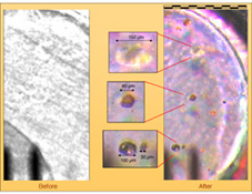
This mosaic of four side-by-side microscope images (one a color composite) was acquired by the Optical Microscope, a part of the Microscopy, Electrochemistry, and Conductivity Analyzer (MECA) instrument suite on NASA's Phoenix Mars Lander. Taken on the ninth Martian day of the mission, or Sol 9 (June 3, 2008), the image shows a 3 millimeter (0.12 inch) diameter silicone target after it has been exposed to dust kicked up by the landing. It is the highest resolution image of dust and sand ever acquired on Mars. The silicone substrate provides a sticky surface for holding the particles to be examined by the microscope.
Martian Particles on Microscope's Silicone Substrate
In figure 1, the particles are on a silcone substrate target 3 millimeters (0.12 inch) in diameter, which provides a sticky surface for holding the particles while the microscope images them. Blow-ups of four of the larger particles are shown in the center. These particles range in size from about 30 microns to 150 microns (from about one one-thousandth of an inch to six one-thousandths of an inch).
Possible Nature of Particles Viewed by Mars Lander's Optical Microscope
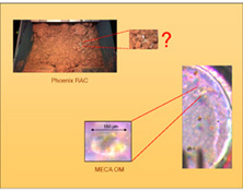
In figure 2, the color composite on the right was acquired to examine dust that had fallen onto an exposed surface. The translucent particle highlighted at bottom center is of comparable size to white particles in a Martian soil sample (upper pictures) seen two sols earlier inside the scoop of Phoenix's Robotic Arm as imaged by the lander's Robotic Arm Camera. The white particles may be examples of the abundant salts that have been found in the Martian soil by previous missions. Further investigations will be needed to determine the white material's composition and whether translucent particles like the one in this microscopic image are found in Martian soil samples.
Scale of Phoenix Optical Microscope Images
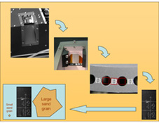
This set of pictures in figure 3 gives context for the size of individual images from the Optical Microscope on NASA's Mars Phoenix Lander.
The picture in the upper left was taken on Mars by the Surface Stereo Imager on Phoenix. It shows a portion of the microscope's sample stage exposed to accept a sample. In this case, the sample was of dust kicked up by the spacecraft thrusters during landers. Later samples will include soil delivered by the Robotic Arm.
The other pictures were taken on Earth. They show close-ups of circular substrates on which the microscopic samples rest when the microscope images them. Each circular substrate target is 3 millimeters (about one-tenth of an inch) in diameter. Each image taken by the microscope covers and area 2 millimeters by 1 millimeter (0.08 inch by 0.04 inch), the size of a large grain of sand.
The Phoenix Mission is led by the University of Arizona, Tucson, on behalf of NASA. Project management of the mission is by NASA's Jet Propulsion Laboratory, Pasadena, Calif. Spacecraft development is by Lockheed Martin Space Systems, Denver.
Photojournal Note: As planned, the Phoenix lander, which landed May 25, 2008 23:53 UTC, ended communications in November 2008, about six months after landing, when its solar panels ceased operating in the dark Martian winter.
